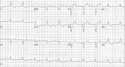
QRS > 0.12sec
Look at V1:
- Terminal R = RBBB as excitation spreading from left to right
- Terminal S = LBBB as excitation spreading away from right
Confirm I:(& aVL V5&6)
- Terminal S = RBBB as excitation going away from left side
- Terminal R = LBBB as excitation heading towards left
The above equates to pattern recognition of MaRrow/ WiLliam in V1-6.
With LBBB associated ST/T opposite to QRS, poor R progression in V1-6, RS in V5,6 left axis deviation.
Diagnosing AMI in LBBB
The Sgarbossa criteria only apply in LBBB (see rules above)
In true LBBB, there must not be any Q wave in the lateral leads

Sgarbossa criteria
Of acute MI with LBBB (any of following)
- ST elevation ≥ 1mm concordant with QRS
- ST depression ≥ 1mm in V1-3
- ST elevation ≥ 5mm discordant with QRS
Hemi-blocks – QRS ≤ 0.12sec
Left Anterior Superior Fascicular Block:
Left ventricle stimulated from LPIF so direction of excitation is Inferior to Superior and Right to Left so
- left axis deviation < -45°
- R in I > R in II or III
- qR in aVL
- deep S in II III aVF
Left Posterior Inferior Fascicular Block: (Rare in isolation as anatomically less discrete than LASF – often accompanies RBB as bifascicular block, or in association with a significant degree of ischaemic damage.)
Left ventricle stimulated from Superior to Inferior and Left to Right:
- Right axis deviation > 110°
- Small r & deep S in I
- R in III > II
- qR in III
Trifasicular Block
Indicating advanced disease:
- RBB and LASF with 1st degree AV block
- RBB and LPIF with 1st degree AV block
- LBB with 1st degree AV block
- Alternating RBB and LBB
Risk of sudden progression to complete heart block or sudden death low unless associated with acute MI or symptomatic bradycardic episodes - prophylactic pacing for these patients.