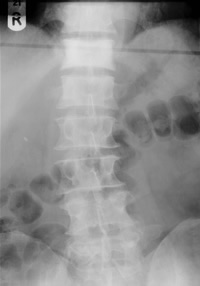
Back pain may be due to 1. mechanical back pain 2. neurogenic pain, 3. pathological pain and 4. inorganic pain
Mechanical back pain
- Is due to sprain of the spinal ligaments or arthritis in the facet joints
- Confined to the back (occasionally referred into the upper leg)
- Exacerbated by movement
- Relieved by rest
- If pain at night, it is when they roll over
- PMHx of similar problem / previous injury making back more susceptible
Red flags
- Age >55 or <19
- Night pain
- Thoracic back pain
- Bilateral neuro. symptoms
- Sphincter symptoms
- Immunosuppression (or IVDU)
- Hx malignancy or Wt. loss
- Fever or recent infection
- Coagulopathy
Neurogenic pain
- Nerve root is irritated by prolapsing lumbar disc, # or osteophyte
- Pain and paraesthesia
- Distribution consistent with that nerve root.
- Paraesthesia is in the same distribution:
- L4: pain extends into the medial malleolus
- L5: into the lateral malleolus and big toe
- S1: pain into the lateral border of the foot and little toe
- Paraesthesia is in the same distribution:
- Worsening disease results in loss of sensation / power / reflex.
- Distribution is characteristic for each nerve root
Pathological pain
- Nasty, unpleasant pain at rest
- Nocturnal pain prominent
- Due either to malignant disease or infection
- If neurological symptoms,may initially seem fluctuant / inconsistent
- Neurological dysfunction can rapidly result if it is not resolved
- Past History:
- Thoracotomy, Mastectomy, Prostatectomy

Exam - non-traumatic back pain
Examine spine
- History, history, history
- Look (scoliosis, loss of lordosis, spasm)
- Feel (tender vertebra/rib, step betw spinous process)
- Move - limitation and pain
Please remember AAA and retroperitoneal pathology (pyelonephritsis, haematoma, pancreatitis) may present as back pain and consider appropriate tests (US AA in all older patients with back pain).
Beware stiffness in young people associated with sclerosis of SI joints. Check ESR.
Neurology
- Sensation (perianal pin prick)
- Coordination (CNS/cord lesion)
- Power and reflexes
- SLR/sciatic stretch test
- Femoral nerve stretch test
- FAIR (hip flex/add/int. rotation) - piriformis pathology
Sciatic (L5 root) vs other pathology
Sciatic nerve (L5):
Weak ankle dorsiflexion/plantarflexion, foot inversion/eversion; absent ankle jerk. May have weak knee flexion. Weak hip abduction if L5 lesion (sup gluteal nerve).
Piriformis syndrome:
Trigger point in buttock. Treat with physio.
Iliotibial band syndrome:
Recent Δ activity or footwear (inversion). Trigger with Ober's test and weak popliteus.
Peroneal nerve pathology:
Hx Local trauma (fibular neck), tender peroneal nerve, weak ankle dorsiflexion and foot eversion, but strong ankle plantarflexion and foot inversion, normal AJ, paraesthesia dorsum of the foot only.
Cortical lesion:
+ve Babinski.
Cauda equina:
See below. Bilateral symptons ± sphincter.
Central "disc" prolapse - cauda equina
- Is a medical emergency requiring urgent MRI
- Bilateral radicular pain
- Δ perianal (pin prick) sensation or genital dysaesthesia
- Bilateral motor weakness (esp. knee extension, anle eversion, foot dosiflexion)
- Lax anal sphincter (late, unreliable sign)
- New (<14/7) micturition difficulty (post void residual vol. >200ml)
- If unable to micturate, check bladder vol:
- If > 600ml, catheterise (to avoid distension injury)
- Document if sensate and catheter tug
- Contact your senior now
X-rays required if:
- Direct trauma
- Relevant Hx
- Patients with neurological signs
- Unable to stand
- Elderly
- For ortho. opinion (they'll ask)
Management
Non-Pharmacological Interventions
- Patient self management Advice sheet - Print
- Physiotherapy: In patient Physiotherapy in CDU in severe cases, otherwise outpatient physiotherapy(mainstay of treatment)
- Psychological Therapy
- Consider cognitive-behavioural therapy (CBT) by psychologis
- Return-to-Work Programs: encourage return to work and daily activities
Pharmacological Management of Low Back Pain
- NSAIDs : Use the lowest effective dose and provide gastroprotective treatment if needed while considering risk factors. (See "Special Populations")
- Weak Opioids: considered ONLY if NSAIDs contraindicated or ineffective
- Paracetamol : Do not offer paracetamol alone for low back pain management
- Other Medications: Do not offer SSRIs, SNRIs, tricyclic antidepressants, gabapentinoids, or antiepileptics for low back pain
- Lidocaine (local anaesthetic) patch topical
Pain clinic referral to be arranged by GP in cases of unsuccessful pain management with above therapies
Special (elderly > 70 yo) populations:
- Caution with NSAIDS due to GI and Renal side effects
- MST/Oramorph (Oral Morphine) is the preferred analgesic
NSAIDS in CUH
- Pharmacology section
- Ibuprofen 400mg q8h PO
- Naproxen/Esomeprazole 500/20mg (Vimovo) q12h PO
- Diclofenac 75mg (Difene) q12h PO
- Diclofenac 100mg q16h PR (only in ED, in-patient, or CDU admission)
- Parecoxib 40mg q12h IV